

超聲引導下腎活檢
超聲引導腎活檢或經皮腎活檢(PRB)是一項技術,其中包括取一小塊腎臟組織進行顯微鏡檢查。
哪種超聲掃描儀用於腎臟活檢?
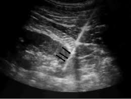
隨著 凸 3.5 MHz換能器的超聲波 西弗特拉斯-5.21,醫生會定位天然腎臟的下極或腎臟同種異體移植的上極,並評估從皮膚表面到活檢點的距離。
然後,醫生應在針將要進入的皮膚表面上做一個標記。 皮, 皮下, and peri-renal tissues penetrates with local anesthetic using ultrasonic guidance, verifying sufficient local anesthesia along the intended biopsy pathway.
To make a smooth passage of the biopsy needle, the doctor should make a small cut through the weal. The biopsy needle is then guided through the skin incision, and then under real-time ultrasonic guidance across the bottom pole of the kidney or the upper pole of the renal allograft.
然後,當看到針尖滲入腎囊時,停止針的前進。 然後開槍,然後立即將套管推過管心針並獲得腎實質的核心。
After the procedure, the kidney can be scanned to evaluate for the existence of hematoma or active bleeding. A second check with ultrasonogram should be done after 24 hours from the procedure to watch for any peri-renal bleed or hematoma which would have developed later.

[啟動板_反饋]
儘管使用了我們提供的信息,但是醫生,放射科醫生,醫務人員執行了其程序,臨床應用,但本文中包含的信息僅供參考。 對於設備的誤用或設備與本文提到的每種臨床應用或程序的適用性,我們概不負責。
醫生,放射科醫生或醫務人員必須具有適當的培訓和技能,才能對每個超聲掃描儀設備執行此過程。
